


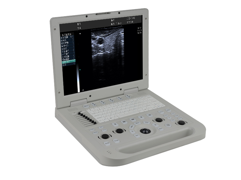
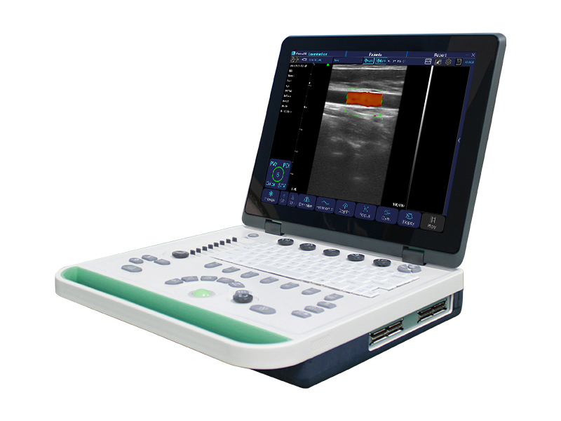
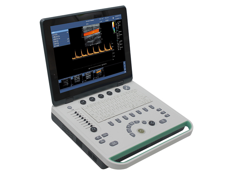
System Imaging Function:
1)Color Doppler Enhancement Technology
2)Two-dimensional grayscale imaging
3)Power Doppler imaging
4)PHI pulse inverse phase tissue harmonic imaging + frequency composite technique
5)With the working mode of spatial composite imaging
6)Linear array probe independent deflection imaging technique
7)Linear trapezoidal spread imaging
8)B/Color/PW trisynchronous technology
9)Multibeam parallel processing
10)Speckle noise suppression technology
11)Convex expansion imaging
12)B-mode image enhancement technique
13)Logiqview
Measurement and analysis:
1)General measurement: Including distance, area, circumference, volume, area ratio, distance ratio, Angle, S/D velocity, time, heart rate, acceleration, etc
2)Obstetrics: Obstetrics supports the measurement of fetal data ≥3 fetals, including fetal weight algorithm, growth curve display, fetal echocardiography measurement (including left ventricular function measurement, left ventricular myocardial weight, etc.); Fetal measurement OB1, OB2, OB3)
3)Blood flow measurement, sampling volume at least 8 levels adjustable
4)Automatic measurement of endovascular media
5)All measurement data Windows are removable
6)Customized comments: Include insert, edit, save, etc
Input/output signal: input: Mquipped with digital signal interface. Output: VGA, s-video,USB, audio interface, network interface.
Connectivity: Medical digital imaging and communications DICOM3.0 interface components.
Support network real-time transmission: can real-time transmission of user data to the server.
Image management and recording device: 500G hard disk Ultrasonic image archiving and medical record management function: complete the storage management and playback storage of patient static image and dynamic image in the host computer
General system function:
1)PC Platform
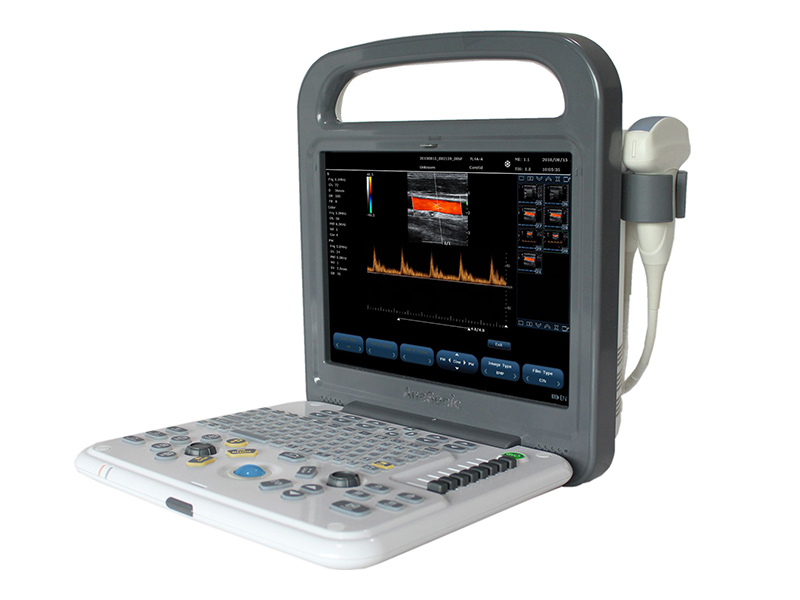
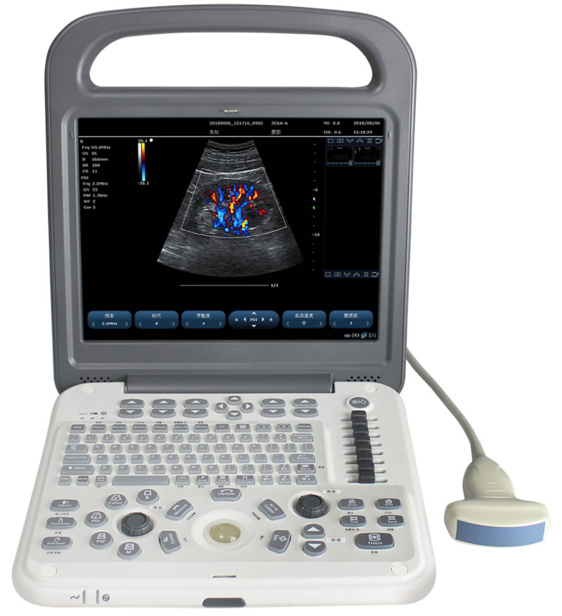
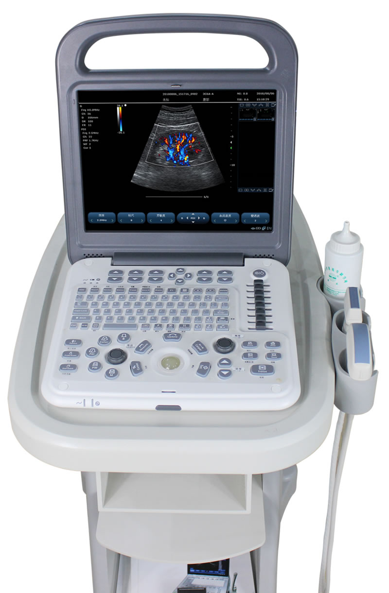
2)Color monitor: 12.1 "high resolution color LCD monitor
3)Probe interface: zero force metal body connector, effectively activated two mutual common interfaces
4)Dual power supply system, built-in large capacity lithium battery, battery power 2 hours duration, and the screen provides power display information
5)Net Weight: 4.5kg
6)Support quick switch function, cold start 39 seconds.
7)Main interface miniatures
8)Full-screen Chinese and English annotation input, Chinese input method 2 kinds, including wubi input method
9)Built-in patient data management station
10)Customized comments: include insert, edit, save, etc
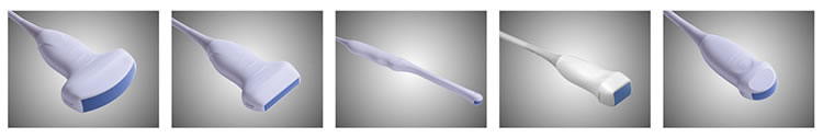
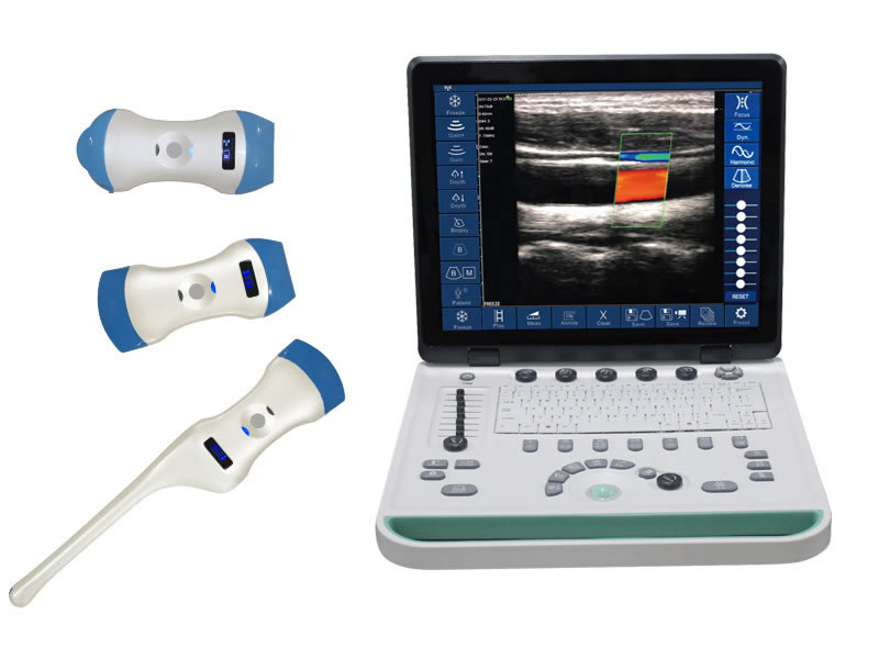
Probe Specifications:
1)2.0-10MHz V¬ariable frequency, frequency range 2.0-10MHz
2)Optional Probes: Convex, Linear, Transvaginal, Phased Array, Microconvex
3)5 kinds of frequencies of each probe, variable fundamental and harmonic frequency
4)Abdomen: 2.5-6.0MHz
5)Superficial:5.0-10MHz
6)Cardiac:2.0-3.5MHz
7)Puncture Guidance: probe puncture guide is optional, puncture line and Angle are adjustable
8)Transvaginal: 5.0-9MHZ
9)B/D Dual-purpose: Linear: B/PW, Convex: B/PW, Sector-Scan: B/PW, Phased Array: B/PW
Main parameters of two-dimensional grayscale imaging:
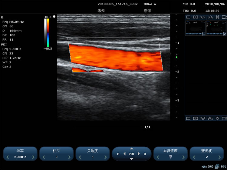
1)Imaging modes including: B Mode, 2B Mode, 4B Mode, M Mode, Color(Color Doppler) Mode, PDI(Power Doppler) Mode, PW Mode, CW Mode, Dissection M, B+C+PW three modes, B+PDI+PW three modes, support single window display, double window real-time display, four windows display
2)Imaging speed: the maximum frame speed is 80 frames per second
3)Beam focus: 4 focal points
4)Real-time amplification (unlimited) and freeze amplification (3 times)
5)2D image gain, continuously adjustable
6)Receiving mode: parallel processing of multi-beam signals
7)The sound power is adjustable by sight
8)Scanning Depth(mm)≥351mm
9)Support Kidney Stone breaking line
10)Sound beamformer: digital sound beamformer, digital full-range dynamic focusing, digital dynamic variable aperture and dynamic trace, and focus position can be adjusted in the whole imaging area
11)Playback: image playback for 48 seconds (Phased Array B mode)
12)False color 7 kinds of color
13)Preset conditions: for different inspection organs, preset the optimal image inspection conditions, reduce the adjustment during operation, and commonly required external adjustment and combination adjustment
14)segment adjustment for 8 segments
15)Sector Scan Angle: 4 grades adjustabl
16)TEI Index
17)2D Modes(B) Phased Array: maximum: ≥6898 frames, Color,PDI maximum: ≥4050 frames
Spectral Doppler technique requirements:
1)Transmission Mode: pulse wave doppler PW, continuous wave doppler CW
2)PW testing range 0~7.5m/s
3)Doppler Frequency: PW frequency
4)Maximum measured velocity: positive or reverse flow velocity of 7.5m/s
5)Doppler Automatic envelope measurement and calculation
6)SV sampling width and location range : width 1–8mm
7)Display Control: reverse display (left/right;Up/down)
8)PW Real-time automatic measurement function
8)Scaleplate ≥16 grades; PRF 0.7kHz-9.3KHz adjustable
10)Playback: automatic playback of movies
Color Doppler technique requirements:
1)The doppler gain is continuously adjustable
2)Color enhancement
3)B+COLOR display on both left and right sides of the same screen
4)Color mode baseline adjustment ±15 grades
Rich data interface for data analysis:
1)VGA interface
2)Printing interface
3)Network interface
4)SVIDEO interface
5)Foot switch interface


 Home
Home Product
 News
News Contact