The Application of Mini Ultrasound in Lung Scanning
- 2023-03-03
- 2027
- Guangzhou Sonostar Technologies Co., Limited
- A Case Study by Zephyr, Pulmonologist at Taipei Veterans General Hospital
An 86-year-old man was sent to our emergency room for shortness of breath. Chest film revealed cardiomegaly and increased opacity over right hemithorax and blunt bilateral costophrenic angles. Sonography was ordered to determine whether the right hemithorax was filled with pleural effusion or it was a consolidation of the right lung.

Chest film showed increased opacity over the right hemithorax.
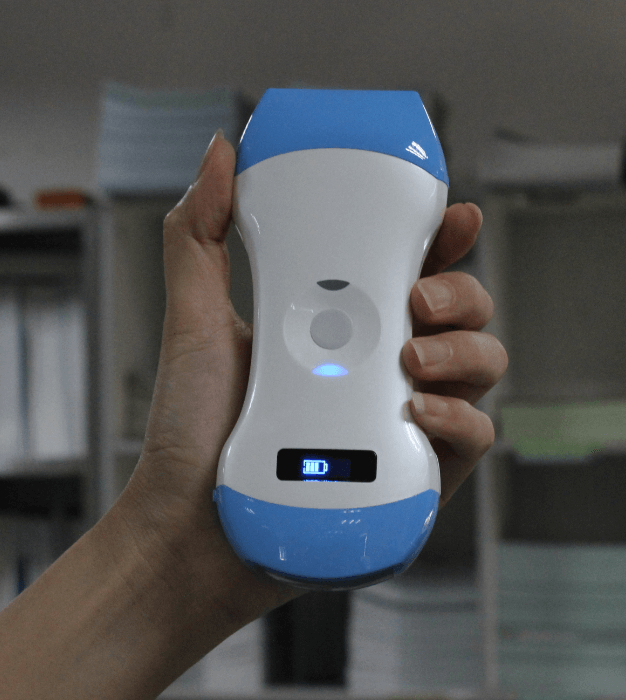
We used Mini Ultrasound during ward rounds and a diagnosis of pleural effusion was made immediately. The video showed the general survey of the patient’s right and left hemithorax. In the general survey, we would start from the side with fewer problems, and place the probe perpendicularly to the ribs at the mid-scapular line and shift downward gradually. When we reach the costophrenic angle, we would rotate the probe parallel to the ribs to see the image without shadows. In the survey of left hemithorax, the bat-wing sign was seen initially that represented the normal lung field, and then hypoechoic pleural effusion appeared. The still image showed a small amount of pleural effusion. on the left side and a large amount of pleural effusion on the left side.
On the contrary, hypoechoic pleural effusion was detected initially on the right side, presenting a large amount of pleural effusion. The collapsed lung is barely seen on the still image due to the pleural effusion is too large.

Small amount pleural effusion with left lower lobe passive atelectasis, diaphragm, and spleen.

A large amount of pleural effusion, diaphragm, and liver.
Under the left decubitus position, after sterilization and local anesthesia, thoracocentesis was performed over the right 6th ICS at the posterior axillary line. Then, a pig-tail catheter, 8 Fr., was inserted over the same route via a two-step method and fixed at 15cm under Mini Ultrasound guidance.

The sonography demonstrated a pigtail catheter inserted into the pleural space filled with pleural effusion and a collapsed lung - Scanned by Mini Ultrasound

Chest film showed a pigtail catheter inserted into the right pleural space.
We checked the pigtail catheter position again after the procedure and the chest film showed the pigtail catheter is in the right position. His breath smoother after then.
Generally, the Mini Ultrasound completed its mission to help clinicians to diagnose pleural effusion and did help we do some sono-guided interventions. The image quality of the convex side is fair as expected. Maybe the image quality would improve in the next vision with more elements.






 网站首页
网站首页 产品中心
产品中心 服务支持
服务支持 联系咨询
联系咨询