Bladder Ultrasound
- 2023-03-03
- 2015
- Guangzhou Sonostar Technologies Co., Limited
Definition
A noninvasive method of assessing bladder volume and other bladder conditions using ultrasonography to determine the amount of urine retention or post-void residual urine.
Purpose
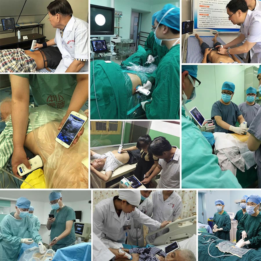
Bladder ultrasound is used in the acute care, rehabilitation, and long-term care environments. It is a noninvasive alternative to bladder palpation and intermittent catheterization used to assess bladder volume, urinary retention, and post-void residual volume in postoperative patients who may have decreased urine output; in patients with urinary tract infections (UTIs), urinary incontinence, enlarged prostate, urethral stricture, neurogenic bladder, and other lower urinary tract dysfunctions; or in patients with spinal cord injuries, stroke, diabetes, and mental handicaps that may reduce the sensation of bladder fullness, thereby interfering with appropriate voiding. Bladder ultrasound may be used in rehabilitation for bladder assessment and training. Bladder ultrasound is used to evaluate bladder function in nursing home residents to monitor for UTIs, urinary incontinence, urinary retention, and bladder dysfunction associated with other medical conditions (e.g., pelvic organ prolapse).
Precautions
There are no contraindications for bladder ultra-sound. However, users should be aware of errors in measurement that may occur. For the most accurate results, patients should be in a relaxed, supine (lying down) position, and the ultrasound scanning head should not be moved during the scan if a portable device is used. Measurements may be distorted in patients with staples or sutures, an indwelling catheter, or scar tissue. Fluid in a pelvic cyst or tumor may be misinterpreted as bladder volume.
Description
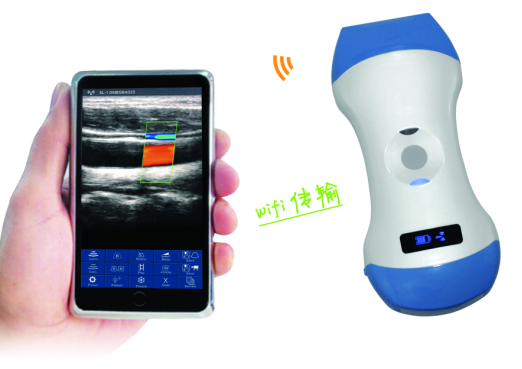
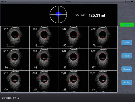
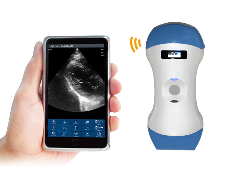
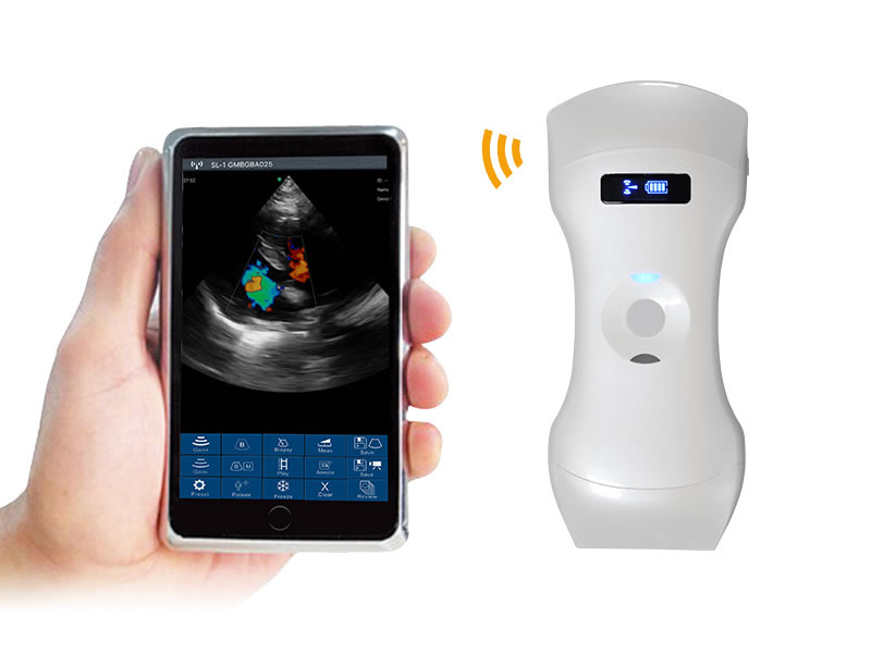
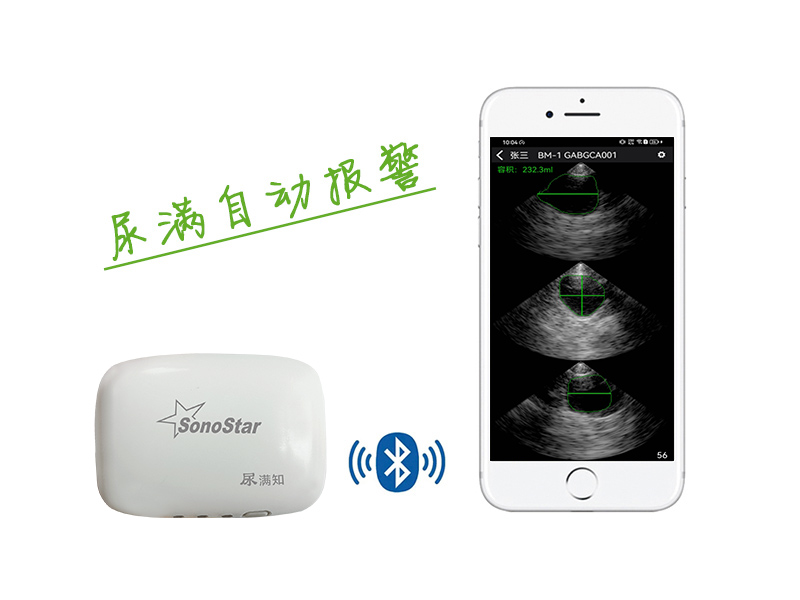
Bladder ultrasound is conducted using a portable, battery-operated ultrasound scanner that consists of a small, handheld unit and an attached ultrasound probe. It may also be performed with a conventional ultrasound unit. The probe, which is placed on the patient's abdomen over the bladder, holds a motorized scanning head with an ultrasonic transducer that transmits sound waves in a fanlike array that are reflected back from the patient's bladder to the transducer. Data from multiple cross-sectional scans of the bladder are then transmitted to a computer in the handheld unit, which automatically calculates bladder volume. The handheld unit also contains an integral digital screen and printer for displaying the bladder volume measurements. The entire scan only takes a minute or two, is noninvasive and painless, and eliminates the discomfort, embarrassment, and risks associated with catheterization.
The bladder ultrasound procedure is also referred to as bladder scanning or the bladderscan, after the brand name of the most widely available portable bladder ultra-sound device. A dedicated portable bladder ultrasound scanner ranges in cost from approximately $6,000 to $10,000.
Although general-purpose ultrasound scanners, such as those used in the radiology department, can be used to measure bladder volume, they may be inconvenient for regular use at the patient's bedside due to their size and are much more expensive than portable units. However, they are often used for bladder ultrasound if there is no portable unit in the facility.
Preparation
Before scanning, the end of the transducer scanning head should be wiped with alcohol and allowed to dry. Ultrasound transmission gel should then be applied to the end of the scanning head. The portable bladder ultra-sound device should be set to either male or female; the male setting should be used for a female patient who has had a hysterectomy. To begin scanning, the tip of the scanning head should be positioned approximately one inch (2.5 cm) superior to the symphysis pubis and pointing toward the bladder. During scanning, the scanning head should be held stationary when obtaining measurements. For obese or elderly patients, abdominal flesh may need to be gently moved to one side while scanning to obtain more accurate results.
Aftercare
After scanning is completed, the ultrasound gel should be wiped from the patient's skin and the scanning head. The scanning head should then be cleaned with alcohol. Bladder volume measurements can then be printed out and attached to the patient's chart. Depending on the bladder volume measured, urethral catheterization is performed to relieve urine retention if the patient cannot void on his/her own. If the patient has an indwelling catheter, the bladder volume measurement may indicate a need for catheter irrigation or checking for catheter blockage. The bladder scan may need to be repeated, depending on the catheterized or voided urine volume obtained.
Complications
There are no complications associated with the bladder ultrasound procedure.
Results
In general, if the bladder volume measured is greater than 300 milliliters, urethral catheterization or patient voiding to relieve urine retention should be performed. Clinical studies have demonstrated that using the bladder ultrasound scan instead of intermittent catheterization to measure urine retention reduces the risk of urinary tract infections.
Health care team roles
In the acute care setting, the nurse and physician are responsible for monitoring urine output in postoperative patients. In the long-term care and rehabilitation settings, the primary responsibility for monitoring urine output and/or bladder function lies with the nursing staff. Device manufacturer representatives provide in-service training to nursing staff on using the bladder ultrasound scanner. Clinical and nurse managers may also implement bladder scanning protocols and results and outcomes tracking, particularly when bladder ultrasound replaces intermittent catheterization protocols with the goal of reducing catheterization-related costs and infections.
KEY TERMS
Post-void residual volume—The amount of urine remaining in the patient's bladder after voiding.
Transducer—The part in the ultrasound scanning head that transmits acoustic energy and converts it into electrical energy to produce image data.
Ultrasonography—An imaging modality that uses sound waves to produce anatomical images and measurements.
Resources
PERIODICALS
Phillips, JoAnne K. "Integrating Bladder Ultrasound into a Urinary Tract Infection-Reduction Project." American Journal of Nursing 100, no. 3 (March 2000, Supplement).<http://www.nursingcenter.com/ce>.
Smith, Diane A. "Gauging Bladder Volume without a Catheter." Nursing 29, no. 12 (December 1999):52-3. <http://www.springnet.com>.
Sulzbach-Hoke, Linda M.; Schanne, Linda C. "Using a Portable Ultrasound Bladder Scanner in the Cardiac Care Unit." Critical Care Nurse 19, no. 6 (December 1999):35-9.
Warner, Amy J.; Phillips, Susan; Riske, Karin; Haubert, Mari-Kay; Lash, Nancy. "Postoperative Bladder Distention: Measurement with Bladder Ultrasonography." Journal of PeriAnesthesia Nursing 15, no. 1 (February 2000):20-5.
Wooldridge, Leslie. "Ultrasound Technology and Bladder Dysfunction." American Journal of Nursing 100, no. 6 (June 2000, Supplement). <http://www.nursingcenter.com/ce>.
ORGANIZATIONS
American Urological Association. 1120 North Charles Street, Baltimore, MD 21201. (410) 727-1100. <http://www.auanet.org>.
Society of Urologic Nurses and Associates. East Holly Avenue Box 56, Pitman, NJ 08071-0056. (856) 256-2335. <http://suna.inurse.com>.
OTHER
Neitzel, Jennifer; Frederickson, Martha; Miller, Elaine Hogan; Cassibo, Lori. The Effects of Bedside Ultrasound Assessment of Bladder Volume vs. Intermittent Catheterization on Postoperative Patient Outcomes. University of Iowa College of Nursing Gerontological Nursing Interventions Research Center. <http://www.nursing.uiowa.edu/gnirc/6thabstracts/ru99-b2.htm>.
Jennifer E. Sisk, M.A.








 网站首页
网站首页 产品中心
产品中心 服务支持
服务支持 联系咨询
联系咨询