


Be Busy in “Fistula”
- 2023-03-03
- 1833
- Guangzhou Sonostar Technologies Co., Limited
No.215 Hospital of Shaanxi Nuclear Industry’s Nephrology Department
Liu Guo
In recent days, I don't know what came over me. I focused on performing several unconventional fistulas. Today, I sorted out and shared them to you.
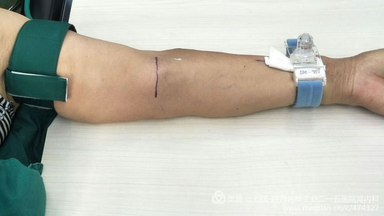
For distance between internal arteriovenous fistula’s elbow vein blood vessel and brachia artery is too close, here is a high incidence area where brachial artery is punctured by mistake.
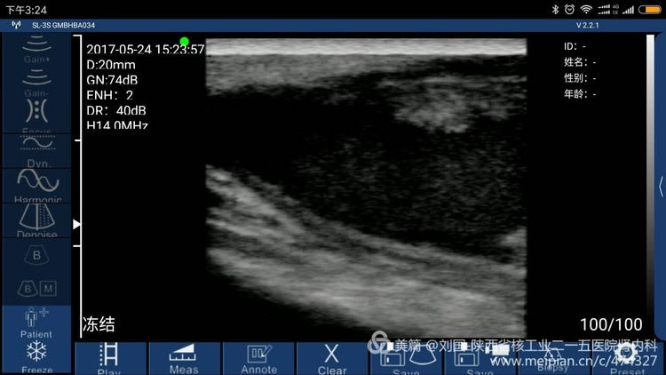
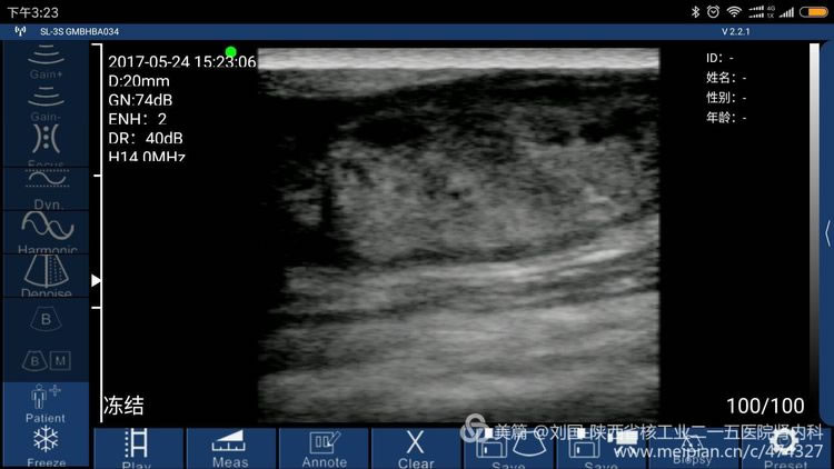
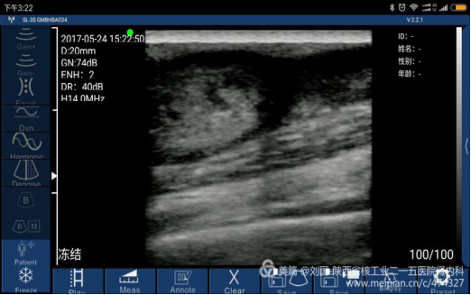
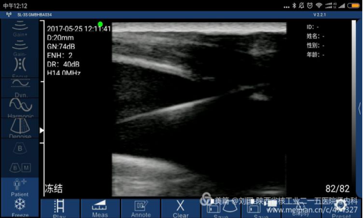
The patient’s barchial artery was punctured in error by a nurse of some hospital’s blood purification center, which caused large area of his arm was bruising and his arm swells, covering up doctors and nurses’ awareness, not to find out mistaken puncture timely and barchial artery’s crevasse, and not to timely oppress the barchial artery effectively. Till the bruising was totally dispersed, the swell faded away, part of the below gradually enlarged to impact the forearm buckling. After he saw a doctor in our hospital, I found I could heard hemokinesis sound. Pseudoaneurysm of elbow brachial artery with 3mm-crevasse was confirmed by ultrasound.
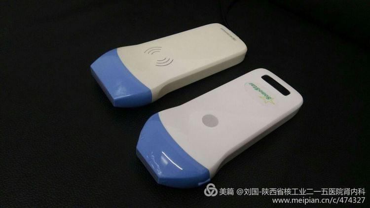
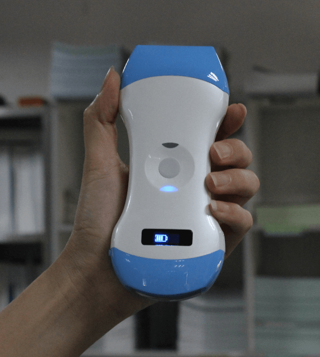
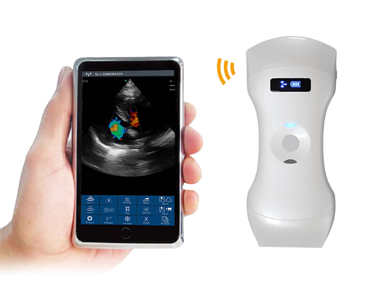
Result checked by Sonostar’s wireless ultrasound, and displayed by my mobile phone-Mi max
Sonostar’s True Features
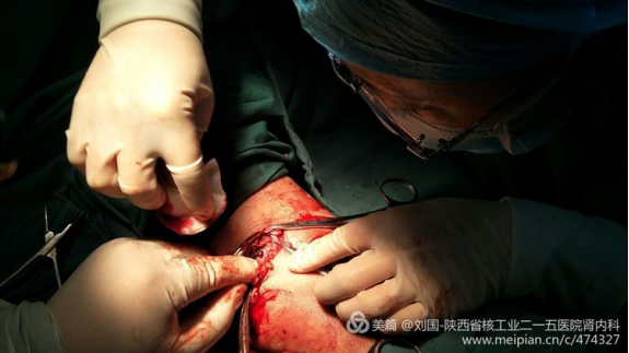
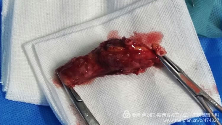
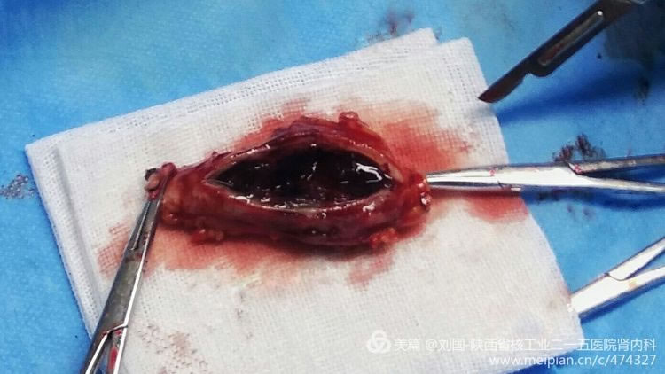
The incision position is located at the right above of brachial artery’s crevasse, the cut direction is along the elbow’s dermatoglyph direction and the cut length is depended on the tumor size.
Cut open the skin, separate the subcutaneous and expose main part of the tumor

Block blood flow at both ends of tumor, cut the tumor, expose the brachial artery’s crevasse, and stitch it. The photo was not clear and could not expose the crevasse and sutured condition.

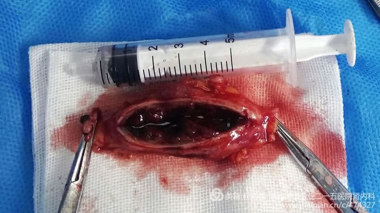
The crevasse has been stitched and the fingers is searching the tumor’s lacunula.
Simply clean mainly including organized blood clot and some tumor tissue in tumor body, and stitch the tumor’s incision.

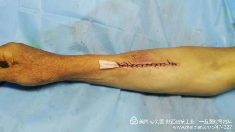
Stitch the skin and wish it to become a thin dermatoglyph.
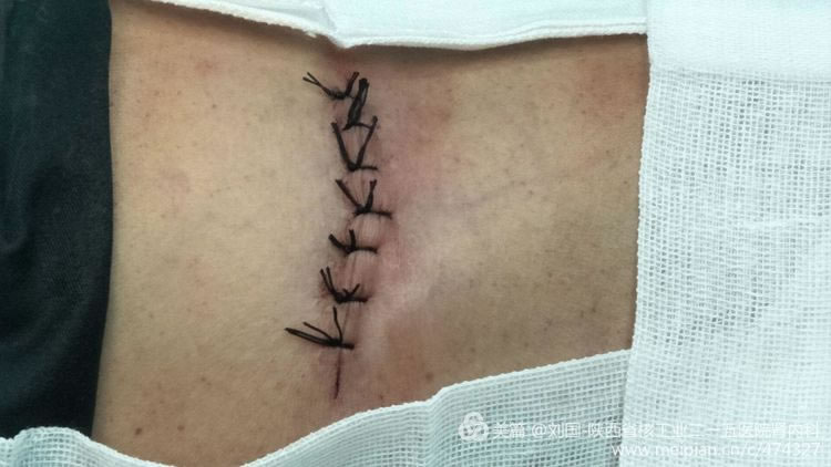
Do not delay and continuously dialyze, adorable!

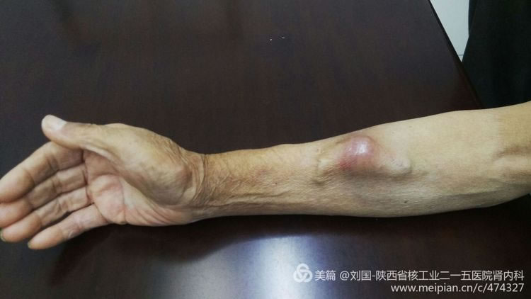
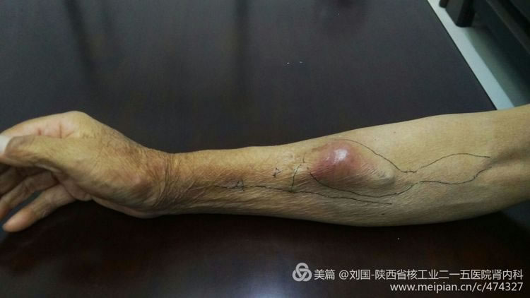
case 2: Excision about Forearm Internal Arteriovenous Fistula Cephalic Vein Tumor-like Expansion and Thrombus to Tumor
The patient’s elbow brachial artery was punctured to cause his local swelling by mistake during hematodialysis when he was in hospital in the first time, which caused. There was a crevasse confirmed by the ultrasound on his brachtial artery, and it was healed after a part of his brachial artery was compressed.
He is hospitalized this time for internal arteriovenous fistula’s cephalic vein tumor-like expansion and thrombegenesis appears in his right forearm, which blocks his cephalic vein from his orificium fistulae to elbow. After his internal fistular was blocked, he performed the internal arteriovenous fistular operation in Xi’an Hospital. When he came to our hospital, the catheterization hemodialysis in his right jugular vein was being executed, part of his right forearm’s orificium fistulae was swelling with severe pain, and his internal thrombus had been organized, which brought much organized thrombus in the cephalic vein from orificium fistulae to elbow without obvious lacuna.
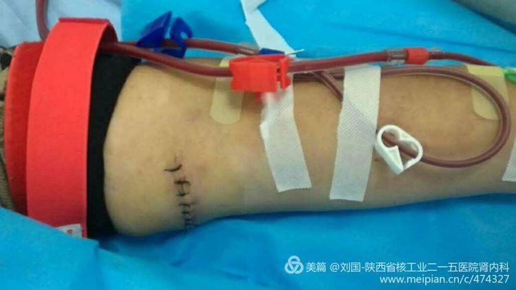
For the compression therapy affects the internal fistula, a intravenous indwelling catheter with Cuff was temporarily put in the patient’s right neck. After the blood purification treatment, his vascular would be repaired to continuously use the internal fistula, and then his intravenous indwelling catheter would removed.
After the brachial artery crevasse was healed, the patient discharged from the hospital to perform maintenance hemodialysis by his right forearm’s internal arteriovenous fistula in other hospital, but did not remove the intravenous indwelling catheter in his right neck.
Preoperative Look
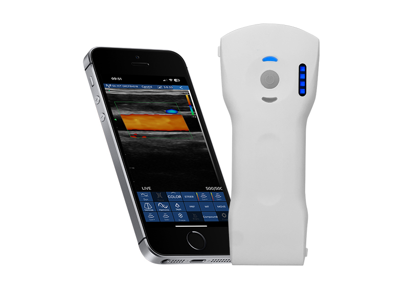
Cephalic vein condition checked by Sonostar’s wireless ultrasound, and displayed by my mobile phone-Mi max
Cut tumor
Open to see
Gesticulate to see the size
One line after operation
Because of facing the popularized internal fistula forming operation, there is no ready-made blood vessel prosthesis at hand, otherwise, the tumor body is cut to put a blood vessel prosthesis, which is perfect
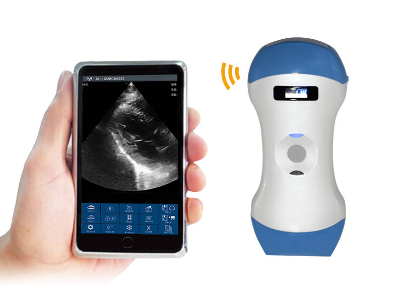
Case 3: Sonostar’s Wireless Ultrasound connected with Mi max by WIFI, synchronously guiding to implant a jugular vein intravenous indwelling catheter with Cuff
Several experiences about using mini wireless ultrasound(Sonostar):
1. It’s better to close the running APP software on mobile phone to confirm the ultrasound software running;
2.For it is connected with the mobile phone as a display by WIFI, image transmission lags. When the display is bad, the operator should slowly move the ultrasound probe, and do not rush, otherwise, a approving image cannot be received.
3.Stumble into the result difference between using iodophor and saline water as coupling agent! Please use saline water!
4.Because there is no color frequency spectrum, the operator cannot judge artery or vein; but the operator cannot judge whether it is artery or vein based on it is compressed or not. The operator should combine physical examination, especial for the peaked patient. I have lessons drawn from my mistakes.
-
 How Mini ultrasound is ultilized in the diagnosis of inguinal lymph node?
 -
 5 Ultrasound Cases in Whole-body Scanning
 -
The application of Mini Ultrasound in detecting gallstone in the gallbladder
-
 Choose the One from All, An Objective Review
 -
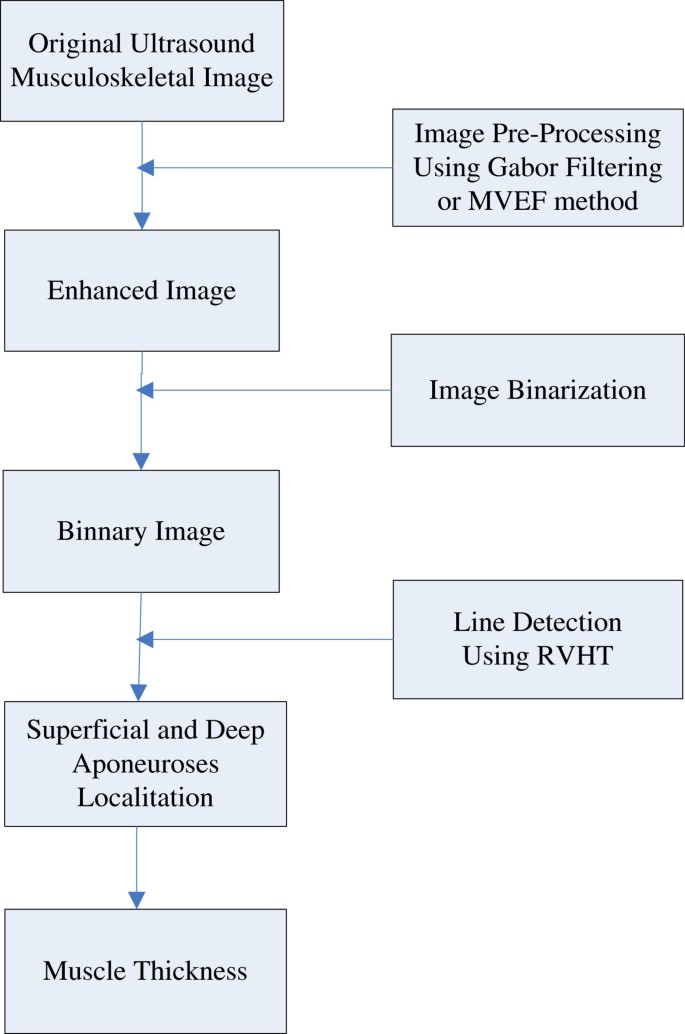 Automatic thickness estimation for skeletal muscle in ultrasonography: evaluation of two enhancement
 -
 Beijing Haidian Hospital’s Nephrological Dept Taking the Lead in Carrying out Wireless Hand Ultrason


 网站首页
网站首页 产品中心
产品中心 服务支持
服务支持 联系咨询
联系咨询