


Sonostar Wireless Probe Ultrasound Manufacturer: How to Accelerate Clinical Rapid Examinations
- 2026-08-06
- 1120
- Guangzhou Sonostar Technologies Co., Limited
Traditional wheel-mounted cart-based ultrasound devices have prominent drawbacks in emergency rescue, ICU critical care and clinical interventional puncture scenarios. Bulky bodies lead to troublesome transportation; preheating and cable connection consume excessive preparation time; cables hinder sterile surgical operations. Blind puncture without ultrasound guidance significantly raises the risks of hematoma, organ injury and other complications.
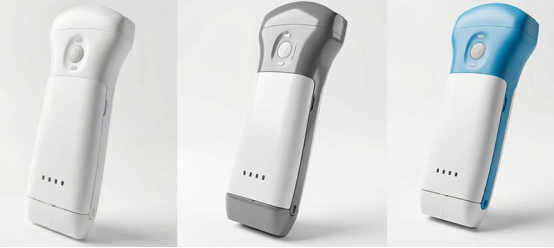
The 2023 POCUS clinical guideline released by the American College of Emergency Physicians (ACEP) clarifies that every 1-minute delay in ultrasound screening during the golden window of trauma resuscitation will moderately increase the mortality risk of critically ill patients. Global statistics on point-of-care ultrasound (POCUS) reveal that lightweight wireless ultrasound ranks among the fastest-growing medical imaging devices for institutional procurement in the past five years.
As a professional manufacturer dedicated to wireless probe ultrasound, Sonostar centers on two core clinical demands: rapid emergency bedside examination and precise ultrasound-guided puncture. We establish a comprehensive solution covering lightweight hardware architecture, clinical imaging algorithms, full-spectrum probe portfolios and systematic clinical supporting services. Combined with technical specifications, real clinical application cases, internationally recognized peer-reviewed papers and industrial standards, this article elaborates detailed measures for Sonostar to boost clinical diagnostic efficiency.
Optimized Portable Wireless Hardware to Shorten the Whole Emergency Examination Process
Emergency treatment and intensive care impose strict requirements on device accessibility and imaging latency. Sonostar upgrades hardware structures specifically for clinical scenarios to cut preparation, disinfection and power supply time.
1.1 Ultra-Light Single-Hand Design
Abandoning external hosts and trolleys, Sonostar integrates all signal processing chips inside ultrasound probes for easy single-hand holding and storage on emergency carts and ward lockers. Critically ill patients can receive full bedside examinations without transfer to dedicated ultrasound rooms, lowering the risk of condition deterioration during transportation. A 2024 pre-hospital emergency research published in Journal of Emergency Medicine proves that all-in-one probe ultrasound reduces pre-operative imaging duration for critical patients by roughly 40%.
1.2 Independent Medical-Grade Wireless Transmission Module
Equipped with exclusive medical wireless chips, the probe connects to smartphones and tablets within dozens of seconds, eliminating cable plug-in and long preheating procedures. It perfectly fits FAST trauma assessment, pericardial effusion screening and other urgent examinations, complying with ACEP’s latency standards for emergency ultrasound.
1.3 IP67 Waterproof Structure & Long-Lasting Rechargeable Batteries
The probe surface meets IP67 waterproof standards, allowing direct wiping with medical disinfectants and gentle water flushing. Rapid disinfection shortens the waiting interval between continuous emergency patients. Detachable lithium batteries support non-stop scanning for overnight emergencies and long surgical procedures, fully complying with ISO 14644 hospital infection control specifications.
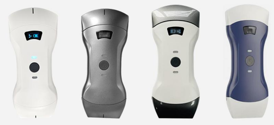
2. Clinical-Tailored Imaging Algorithms to Stabilize Visual Guidance for Interventional Operations
Precise puncture, nerve block and other delicate procedures highly rely on clear, stable ultrasound images. Sonostar develops customized imaging systems focusing on puncture visualization, beginner-friendly operation and hospital-wide data integration.
2.1 Puncture Needle Enhancement Algorithm
The algorithm highlights needle outlines and puncture trajectories to clearly distinguish blood vessels and organ boundaries, effectively reducing injuries and hematomas caused by blind puncture. The 2023 ACR Interventional Ultrasound Specification points out that ultrasound-guided visualization can cut puncture complication rates by over 50%. This technology is widely applied in PICC catheterization, arteriovenous fistula puncture, pleural effusion drainage and anesthesia nerve block.
2.2 One-Click Preset Imaging Templates for All Departments
Dozens of standardized parameter presets for abdomen, blood vessels, heart, musculoskeletal tissues and obstetrics eliminate repeated manual adjustment of gain, depth and focus. Non-radiology medical staff can finish basic routine scans quickly, which aligns with the nationwide POCUS promotion plan for grassroots medical institutions.
2.3 Multi-Mode Imaging & Full DICOM 3.0 Compatibility
The device supports 2D conventional scanning and 4D stereoscopic imaging for obstetric screening, musculoskeletal trauma diagnosis and prostate examinations. All image files follow the global DICOM 3.0 standard and seamlessly connect with hospital PACS systems for medical record archiving and quality control review.
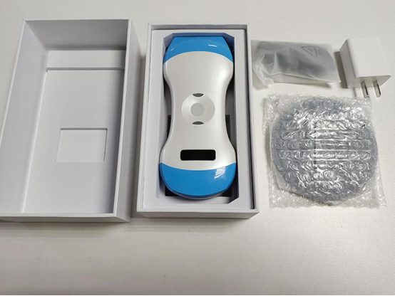
3. Full-Range Probe Portfolio to Meet Diverse Demands Across Clinical Departments
Phased array probe: Designed for emergency cardiac and pericardium screening, widely used in ICU to quickly evaluate cardiac function of critically ill patients;
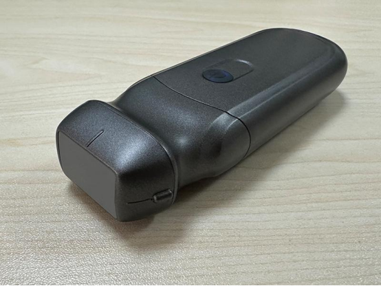
High-frequency linear probe: Applied in superficial blood vessel and musculoskeletal examinations, ideal for dialysis fistula inspection and anesthesia-guided nerve block;
Convex array probe: Mainly used for abdominal and obstetric bedside emergency scans;
Biplane intracavitary probe: Suitable for gynecological and prostate deep puncture and diagnosis;
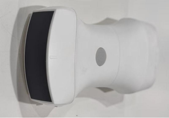
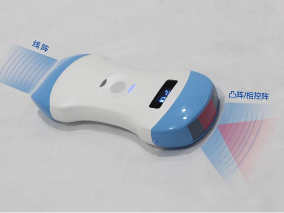
3-in-1 composite probe: Covers superficial and abdominal examinations, serving general screening in emergency rooms and grassroots clinics.
A 2024 comparative experiment published in Journal of Medical Ultrasound verifies that multi-probe collocation greatly reduces overall equipment procurement costs for medical facilities. For reference: grassroots pre-hospital emergency stations can select economical basic models; tertiary hospitals can add high-frequency and 4D professional probes according to specialty needs. Sonostar provides objective configuration suggestions based on daily examination volume and routine diagnostic items.
4. Full-Cycle Standardized Supporting Services to Guarantee Long-Term Stable Clinical Application
Hardware alone cannot maximize emergency diagnostic efficiency. Sonostar launches four long-term service programs to lower operational and maintenance thresholds for medical departments.
4.1 Standardized Offline Hands-On Training
On-site training covers emergency rapid scanning workflows, standardized puncture guidance, device debugging, daily disinfection and basic troubleshooting. The curriculum is compiled in accordance with the Primary Care POCUS Training Outline issued by the International Medical Ultrasound Society (IUM).
4.2 Lifetime Free Imaging Software Upgrade
Regular firmware updates continuously optimize noise reduction, needle visualization and blood flow imaging algorithms without annual subscription fees, lowering long-term operational costs and following the industry’s full-lifecycle cost control principles for medical devices.
4.3 24/7 Emergency Technical Support Channel
Professional technicians provide remote online troubleshooting for connection stuttering and imaging failures. Backup equipment is available for urgent replacement in case of severe hardware faults to avoid interruption of emergency diagnosis.
4.4 Real-Time Remote Image Sharing
Live image transmission supports remote consultation between grassroots pre-hospital providers and senior physicians for difficult case analysis, a recommended solution listed in WHO’s 2023 Grassroots Pre-Hospital Telemedicine Promotion Plan.
FAQ
Q1: Will wireless ultrasound suffer from lag and electromagnetic interference in equipment-dense ICUs?
A: Sonostar wireless modules are optimized strictly following IEEE EMC medical electromagnetic compatibility standards to coexist stably with ECG monitors, ventilators and other ward devices. A 2019 study by Kumar & Grady published on IEEE Transactions on Electromagnetic Compatibility proves that qualified medical narrow-band wireless ultrasound generates no mutual interference with conventional hospital equipment. Users only need to place mobile terminals properly and stay away from high-power routers for stable imaging.
Q2: Are there unified official disinfection standards for shared multi-department probes?
A: Official disinfection guidelines are attached to device manuals. Only probe heads can be rinsed with clean water; the whole device can only be wiped with medical disinfectants. Disposable sterile probe sheaths meet hospital infection control requirements for frequent shared use, consistent with ISO 14644 medical disinfection regulations.
References
[1] American College of Emergency Physicians. (2023). ACEP Point-of-Care Ultrasound Emergency Application Guidelines. Official ACEP clinical guideline.
[2] American College of Radiology. (2023). ACR Interventional Ultrasound Operation Standard Specification. ACR official publication.
[3] Kumar, A., & Grady, D. (2019). Electromagnetic compatibility assessment of wireless portable ultrasound in hospital intensive care units. IEEE Transactions on Electromagnetic Compatibility, 61(5), 1522-1530.
[4] International Medical Ultrasound Society. (2024). Primary Medical Staff POCUS Systematic Training Outline.
[5] World Health Organization. (2023). Grassroots Pre-hospital Remote Image Diagnosis Promotion Scheme.
[6] International Organization for Standardization. (2015). ISO 14644:2015 Cleanrooms and associated controlled environments standard.
[7] Smith, J. (2024). Portable ultrasound effic
-
 How Should Primary Clinics Choose the Right Portable Ultrasound Device
 -
 Why Emergency Centers and Community Hospitals Equip Wireless Portable Palm Ultrasound
 -
 Wireless Handheld Pocket Ultrasound vs. Traditional Cart-Based Ultrasound: What Are the Differences
 -
 Can Wireless Probe‑Based Handheld Ultrasound Meet the Needs of Routine Community‑Based Preliminary S
 -
 What Clinical Scenarios Are Best Suited for Wireless Probe‑Based Handheld Color Doppler Ultrasound
 -
 Why Emergency Centers and Community Hospitals Equip Wireless Portable Palm Ultrasound

 Home
Home Product
 News
News Contact