


Portable Doppler Ultrasound: How It Detects Blood Flow, Modes, and Real Limits
- 2026-09-14
- 878
- Guangzhou Sonostar Technologies Co., Limited
A portable Doppler ultrasound adds one capability a plain B-mode scanner cannot offer: seeing and measuring moving blood. That single difference decides whether a clinician can confirm vessel patency, guide a needle around arteries, or grade flow at the bedside. This guide explains how a pocket-sized device does it, compares the four Doppler modes, maps them to real clinical tasks, and sets out the physical limits every buyer should understand before choosing a probe.
What Is Portable Doppler Ultrasound?
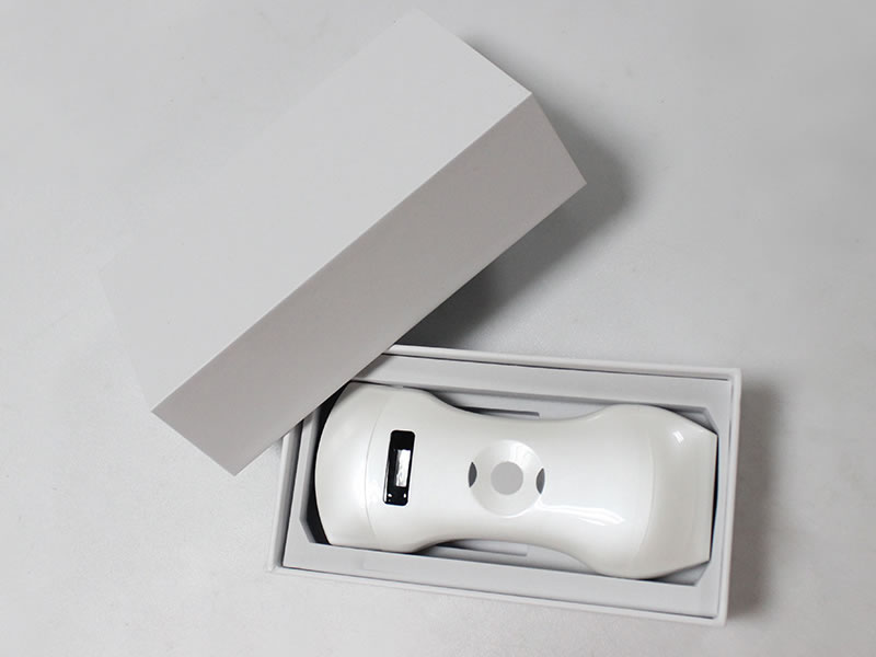
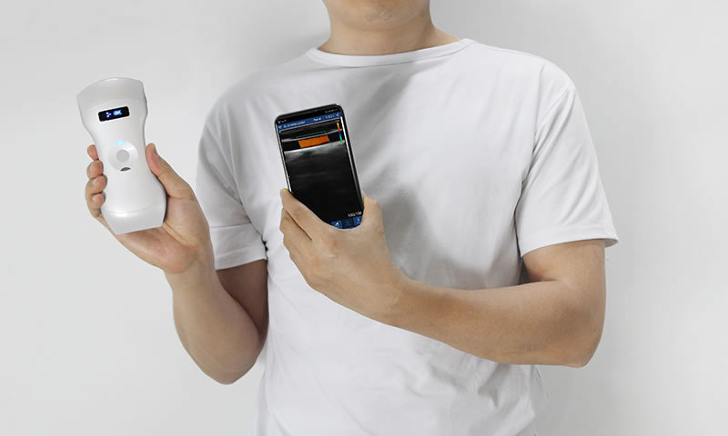
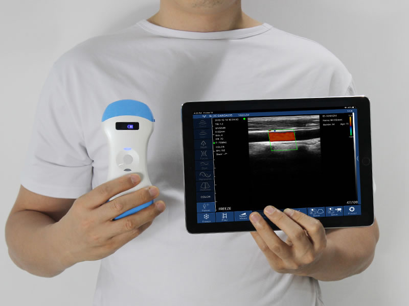
Portable Doppler ultrasound is a compact, often wireless probe that applies the Doppler shift — the frequency change between an emitted pulse and the echo returning from moving red blood cells — to show flow direction, distribution, or velocity on a phone, tablet, or integrated screen. Manufacturers often label these units palm Doppler ultrasound scanners.
Doppler imaging has five recognized forms: continuous-wave (CW), pulsed-wave (PW), color Doppler, power Doppler, and spectral duplex scanning. Most handheld platforms combine B-mode with color, power, and PW spectral Doppler; pure CW remains common in small bedside fetal and vascular flow detectors. The compact format changes the hardware, not the physics.
How Portable Doppler Ultrasound Detects Blood Flow
The probe fires a pulse at a known frequency. Echoes from blood moving toward the probe return at a higher frequency, and echoes from blood moving away return lower; the system encodes the two directions, typically red and blue.
Two physical rules dominate image quality:
Insonation angle. Velocity is reliable only when the beam is close to parallel with flow, and the correction angle should stay at or below 60°. At 90° the cosine is zero, so a perfectly open vessel shows no color — a "pseudo-absence" of flow easily mistaken for occlusion.
Nyquist limit and aliasing. PW and color Doppler sample intermittently, so velocity above half the pulse repetition frequency (PRF) wraps around the baseline or flips color. Fixes are raising PRF, shifting the baseline, lowering transmit frequency, or switching to CW for high-speed jets.
These are properties of ultrasound physics, identical on console systems and pocket probes.

Color, Pulsed-Wave, Power, and CW: Four Modes Compared
| Mode | Output | Range-resolved? | High velocity? | Typical task |
|---|---|---|---|---|
| Color | Direction and distribution over B-mode | Yes | Aliases past Nyquist | Map vessel course, screen patency |
| PW spectral | Velocity waveform at a sample volume | Yes, gated | Aliases past Nyquist | Quantify PSV/EDV, RI/PI |
| Power (PDI) | Flow energy, no direction | Yes | Sensitive to slow flow | Find low-flow/small vessels |
| Continuous-wave | Signal along the whole beam | No | No aliasing, >3–4 m/s | High-speed valvular jets |
B-mode describes the vessel wall and lumen; color shows where and which way blood moves; spectral Doppler quantifies velocity and hemodynamic indices. Each answers a different question, which is why multi-mode palm Doppler scanners outperform single-mode flow detectors.
What Is a Portable Doppler Ultrasound Used For?
Portable color and spectral Doppler move flow assessment out of the vascular lab to wherever the patient is. SonoStar's own vascular application notes map the same tasks seen in daily practice: central and peripheral venous assessment and puncture, arterial assessment, varicose-vein work, stroke screening, peripheral arterial puncture, and carotid atherosclerosis.
Peripheral vessels: patency checks, stenosis screening, DVT triage
Procedure guidance: trace vessels before puncture, keep the needle path clear
Shock and critical care: bedside flow status, pericardial effusion and volume assessment
OB/GYN: fetal heart activity and placental/umbilical flow
Ward follow-up: repeated checks without transporting the patient
| Task | Preferred mode | Frequency band |
|---|---|---|
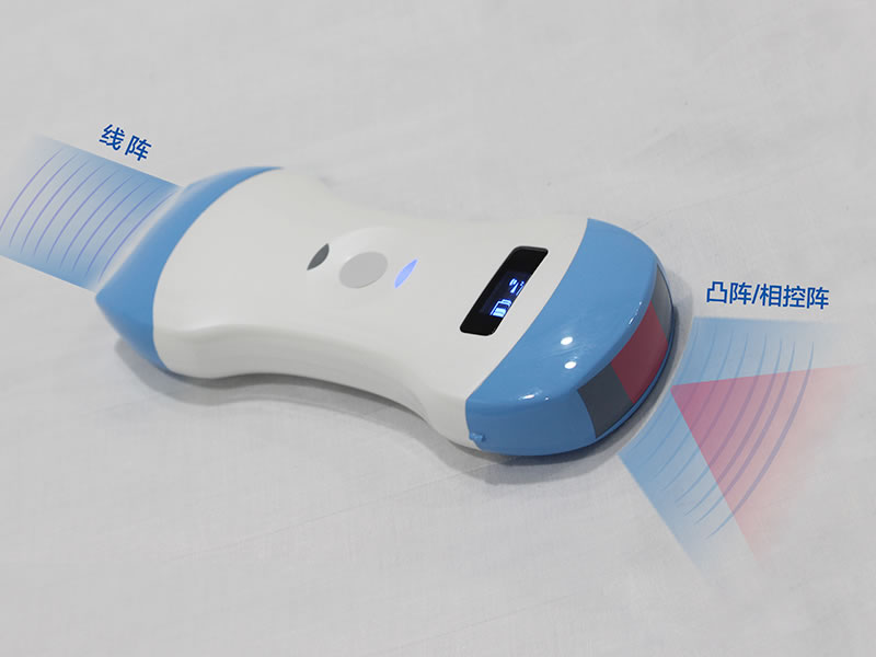
| Superficial/small vessels, nerves | Color + PDI, 20–60 mm | 10–13.2 MHz linear |
| Peripheral vessels, puncture | Color + PW, 20–100 mm | 7.5–10 MHz linear |
| Cardiac / deep vessels | PW spectral, 90–190 mm | 2.2–3.6 MHz phased/convex |
Real Limits of Portable Doppler Ultrasound
Small size does not suspend physics, and four limits recur:
Angle error dominates. Velocities recorded above 60° are unreliable on any device class.
Aliasing in fast flow. Pocket systems offer fewer PRF groups than carts, so high-velocity jets may need CW or a lower-frequency probe.
Wall-filter trade-off. Filters set too high erase genuine low-velocity venous flow; set too low and motion noise floods the spectrum.
Channel count. Fitting beamforming into a ~200 g probe constrains deep, weak-flow sensitivity; complex vascular diagnosis still belongs on a console duplex system.
Operator skill is the other half: gain, sample-volume placement, and angle correction require the same training as a console machine.
Inside a Palm Doppler Scanner: Controls That Matter
The gap between a color-only novelty and a usable duplex device sits in its controls. Three wireless designs from SonoStar's current range illustrate what to ask for:
8C convex palm Doppler scanner — 192 elements, 64 channels, dynamic range adjustable in steps from 40 to 110, suited to deeper abdominal and pelvic flow.
8X linear probe — built around accurate in-plane/out-of-plane puncture guidance, with the same 40–110 dynamic-range steps and a 2200 mAh battery for long procedures.
4CPLT interchangeable-probe wireless Doppler scanner — presets for vessels, carotid, blood flow and cardiac work, with automatic vascular measurement.
Across these designs, the settings that actually change a reading are PRF groups, wall-filter level, PW sample volume (1/2/5 mm on SonoStar linear probes), steerable color box, and angle correction across ±85°. Request these numbers directly — the label "color Doppler" alone does not guarantee quantitative spectral Doppler.
Frequently Asked Questions
Can a portable Doppler ultrasound measure velocity quantitatively?Yes, with PW spectral Doppler: a range-gated sample volume produces PSV/EDV waveforms and indices such as RI/PI. Color-only units show direction and distribution but do not replace spectral measurement.
What is the difference between color and power Doppler on a handheld probe?Color encodes direction and velocity by shade; power Doppler shows flow energy without direction and is more sensitive to slow or tiny vessels.
Why does flow vanish when the vessel is clear in B-mode?The beam is usually near 90° to flow, where the Doppler shift is zero. Heel-toe the probe or steer the color box under 60° before calling a vessel occluded.
Can it replace a cart-based vascular ultrasound?No. It handles focused screening, guidance, and follow-up; definitive grading of complex disease and high-velocity jets beyond its PRF still require a console duplex system.
Which frequency fits superficial versus deep vessels?10–13.2 MHz for 20–60 mm superficial structures, 7.5–10 MHz for peripheral vessels at 20–100 mm, and a 2.2–3.6 MHz phased or convex probe for cardiac and deep vessels.

Conclusion
Portable Doppler ultrasound turns a pocket probe into a flow-assessment tool, but its value depends on matching mode and frequency to the task: color for mapping, PW for quantification, power for slow flow, CW for high velocity. Keep angle discipline and an honest escalation path to console imaging, and compare devices by their actual spectral controls rather than the word "color" on a spec sheet. Review the palm Doppler scanner lineup and request a configuration matched to the vessel depths and procedures your team handles daily.
References
Vascular Ultrasonography. NCBI Bookshelf NBK567746 (StatPearls).
Doppler Ultrasound — five Doppler methods, PMC4923974.
High-quality color Doppler of vascular access. J Vascular Access. 2021 (PMC8607315).
Kremkau FW. Sonography: Principles and Instruments. Elsevier.
Nielsen MB, et al. EFSUMB Position Paper on Handheld Ultrasound. Ultraschall in der Medizin. 2019;40:e1.




 Home
Home Product
 News
News Contact