

Handheld Doppler Ultrasound Scanner: Which Mode to Use?
- 2026-09-20
- 868
- Guangzhou Sonostar Technologies Co., Limited
Handheld probes put B-mode, color, power Doppler and pulsed wave on the same screen, and it is easy to open every button at once. You do not need to memorize the physics. For most daily work, you simply match the question you are asking to the right mode — and the right probe depth.
The table below maps a practical task to the mode that answers it, what you actually see on screen, and which SonoStar probe covers it.
| What you need to do | Mode | What appears on screen | SonoStar probe |
|---|---|---|---|
| See structure, locate anatomy | B-mode | Black-and-white anatomy image | Whole range |
| See which way blood flows | Color Doppler | Red/blue overlay on vessels | 9N, 8C |
| See slow or tiny flow | Power Doppler (PDI) | Flow strength, no direction | 9N, 8C |
| Measure a specific flow speed | Pulsed Wave (PW) | Spectral trace + velocity numbers | 9N, 8C (8C has auto vascular measurement) |
| Superficial vessels, small parts | High-frequency linear | Near-field detail | 9N (10/13.2 MHz, 20–60 mm) |
| Abdomen, pelvis, deep views | Low-frequency convex | Up to 305 mm depth | 8C (3.2/5.0 MHz) |
The workflow is usually the same: start in B-mode to find the structure and steady the probe; switch to color when you need flow direction; move to power Doppler when the flow is slow or tiny; open PW only when you need an actual velocity number. Leaving color and PW on all the time makes the image busy and drains the battery.
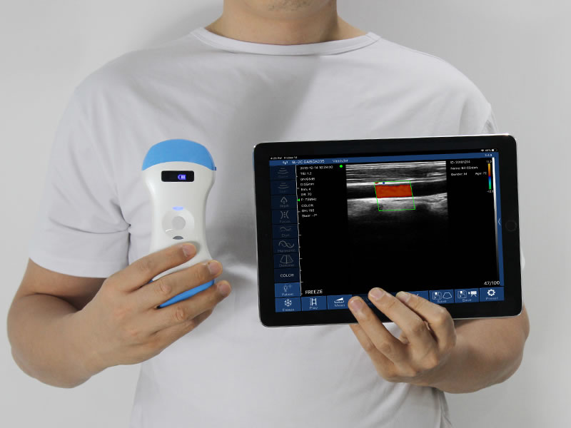
Two points worth clarifying. On color Doppler, red and blue only mean flow toward or away from the probe — they do not by themselves mark an artery versus a vein, so read them together with the B-mode image. Power Doppler does not show direction; its strength is amplifying weak, slow flow. PW samples one point inside the vessel, so a misplaced gate or poor angle gives wrong numbers. SonoStar's convex 8C adds automatic vascular measurement on the PW trace, reading diameter and peak/end-diastolic velocity directly instead of asking the user to caliper by hand.
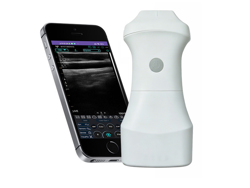
Battery is a practical limit: color and PW draw more than B-mode. For long clinics or outreach, swappable-battery probes that ship with two cells let you swap in two minutes instead of stopping the shift. Scans save as JPG, video loop or DICOM for review and PACS integration.
Match the mode to the question — color for direction, power Doppler for slow flow, PW for a number — and stay in B-mode the rest of the time. That is how a handheld Doppler ultrasound scanner stays clear rather than busy.




 Home
Home Product
 News
News Contact