


Types of Ultrasound Probes: Convex vs Linear vs Phased Array
- 2026-10-14
- 52
- Guangzhou Sonostar Technologies Co., Limited
Before price, brand, battery life or screen size, one decision determines what an ultrasound exam can show: which transducer array touches the patient. The array sets the image shape, how deep the beam reaches, how fine the detail is, and whether the probe can physically get to the target at all. A probe designed for the abdomen cannot produce a useful image of a radial artery, and the reverse is equally true — this is geometry, not marketing.
This guide walks through each main type of ultrasound probe, what its image actually looks like, the frequency and depth it covers, and where it belongs clinically. Every array described here exists in wireless handheld form, so the array choice and the form-factor choice can be made independently.

The Four Core Array Designs
1. Convex (curvilinear) array
Elements sit along a curved face, so the beam fans out into a wide field — wider than the probe's own footprint. On screen this looks like a broad sector, narrow at the top and flaring toward depth.
Typical frequency: roughly 1–8 MHz
Typical depth: to 300 mm or more
Image: wide fan
Used for: abdomen (liver, gallbladder, kidneys, aorta), pelvis, obstetric scanning, FAST trauma views, bladder volume
Limitation: the curved, larger face needs room; it does not fit between ribs or into small superficial areas
When automatic bladder volume is the entire purpose, the convex principle is taken one step further in a dedicated wireless bladder scanner, which measures volume automatically rather than requiring manual calipers.
2. Linear array
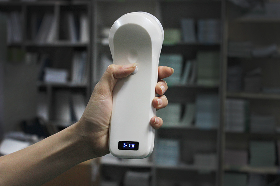
Elements sit in a straight line, sending beams in parallel to produce a rectangular image with no flare. The footprint is flat and wide, which also makes it stable to hold — useful for procedures.
Typical frequency: roughly 5–15 MHz
Typical depth: 20–100 mm
Image: rectangle
Used for: vascular access and vein mapping, arterial assessment, thyroid, breast, scrotum, tendons, muscle, nerves, superficial masses, procedural needle guidance
Limitation: deliberately shallow; it will not reach abdominal depth
3. Phased array
A small cluster of elements pulses with precisely controlled delays, steering and focusing the beam electronically. The result is a sector (pie-shaped) image from a very small contact face — small enough to slide between ribs.
Typical frequency: roughly 1–5 MHz
Typical depth: 90–240 mm depending on model
Image: sector / pie shape
Used for: cardiac windows, pericardial fluid, lung (pleural sliding, B-lines), focused abdominal views, procedural guidance through narrow windows
Limitation: image quality at very shallow depth is not what a high-frequency linear array delivers
Cardiac work needs velocity estimates, so confirm the model supports colour plus PW and CW spectral Doppler. See the full phased array handheld ultrasound guide for the detailed comparison.
SonoStar 5PA: 128 elements / 32 channels, central 2.8 MHz (cardiac harmonic 3.6 MHz), 80°, depth 90 / 160 / 200 / 240 mm, 15–20 frames/sec, ~1 h, 140 × 35 × 25 mm, 120 g, includes CW Doppler.
4. Endocavity array (transvaginal / transrectal)
A curved or micro-convex array mounted on a slim handle, designed to enter a body cavity. Placing the transducer millimetres from the target allows a much higher frequency than an abdominal scan, so resolution improves dramatically.
Typical frequency: roughly 5–8 MHz
Typical depth: 30–120 mm
Image: wide sector or micro-convex fan
Used for: gynaecology and early pregnancy monitoring, pelvic assessment, prostate assessment, follicular monitoring, guidance for oocyte retrieval or prostate biopsy
Note: contact with mucous membranes places these probes in the "semi-critical" category — high-level disinfection between patients plus a sterile probe cover is standard practice

Derivatives Worth Knowing
Micro-convex array
A compromise design: a small curved footprint that still produces a wide field. It reaches places a full-size convex probe cannot — paediatric intercostal windows, small joints, neonatal scanning, small-animal veterinary work — without giving up as much depth as a high-frequency linear array would.
Biplane rectal array
A T-shaped head carries both a convex array and a linear array, giving two perpendicular planes from a single position. Two planes simultaneously means a lesion or needle path can be localised in both axes without moving the probe — which is why the design is used in prostate assessment and anorectal work.
Ultra-high-frequency linear array
Operating in the 16–22 MHz range, this variant trades every millimetre of depth for near-field resolution. It is used for facial aesthetics and vascular mapping, small-part imaging, dermatology, and nerve work where structures sit within a centimetre of the skin.
Ultra-wide linear array
A wider field of view than a standard linear head, at linear-array frequencies. Useful where a larger superficial area needs one pass — chest wall, abdominal wall, extended MSK scanning.
Multi-array probes: 2-in-1, 3-in-1 and interchangeable heads
Rather than carrying separate probes, several arrays can be combined in one housing and switched electronically, or mounted as swappable heads on a shared body.
Two-in-one designs typically pair convex + linear, or linear + phased.
Three-in-one designs house convex + linear + phased in one unit — see the full 3-in-1 ultrasound probe comparison.
Interchangeable-head systems use one body with magnetic hot-swap heads, so the host costs once and the array set grows later.
Types of Ultrasound Probe: Master Comparison
| Probe type | Image shape | Footprint | Typical frequency | Typical depth | Best for |
|---|---|---|---|---|---|
| Convex (curvilinear) | Wide fan | Curved, medium-large | 1–8 MHz | to 300+ mm | Abdomen, pelvis, OB, FAST |
| Linear | Rectangle | Flat, wide | 5–15 MHz | 20–100 mm | Vascular access, thyroid, breast, MSK, nerves |
| Phased array | Sector (pie) | Very small | 1–5 MHz | 90–240 mm | Cardiac, lung, intercostal views |
| Endocavity | Wide sector | Slim handle | 5–8 MHz | 30–120 mm | Pelvic / early pregnancy, prostate |
| Micro-convex | Compact fan | Small curved | 3–8 MHz | 60–160 mm | Paediatric, small joints, vet |
| Biplane rectal | Fan + rectangle, perpendicular | T-shaped head | 6–8 MHz | 40–100 mm | Prostate, anorectal, biopsy guidance |
| Ultra-high-frequency linear | Shallow rectangle | Small-mid linear | 16–22 MHz | 10–40 mm | Aesthetics, superficial vessels, nerves |
| Multi-array (2-in-1 / 3-in-1) | Depends on selected array | Combined | Per array | Per array | Generalists covering multiple regions |
How to Choose: Array First, Everything Else Second
Name the target depth. Under roughly 30 mm → linear or ultra-high-frequency. Roughly 30–120 mm and accessed internally → endocavity or micro-convex. Beyond roughly 90 mm → convex or phased.
Name the access route. Between ribs, subxiphoid, suprasternal → phased array only. Through a body cavity → endocavity. Flat skin surface → linear or convex.
Count the body regions you cover in a day. One region → a dedicated single-array probe gives the best result per dollar. Two or three regions → a combination probe avoids carrying and maintaining three devices.
Check Doppler requirements. Colour Doppler for flow and orientation; PW and CW spectral Doppler whenever velocity or pressure gradients matter — mainly cardiac and some vascular work.
Check needle-guidance features. In-plane and out-of-plane guide lines plus automatic vessel measurement turn a linear or combination probe into an intervention tool.
Then — and only then — optimise form factor. Wireless handheld, cart-based or laptop-style is a workflow question, independent of the array decision.
Specialty Starting Points
| Specialty / setting | Primary array | Often added second |
|---|---|---|
| Emergency medicine | Convex (FAST, abdominal) | Phased (cardiac) + linear (vascular access) |
| Internal medicine / primary care | Three-in-one | — |
| Cardiology | Phased array | — |
| Vascular access / anaesthesia | Linear | Phased (cardiac assessment before blocks) |
| OB/GYN | Convex (transabdominal) | Endocavity (transvaginal) |
| Urology | Biplane rectal | Convex (renal, bladder) |
| MSK / sports medicine / physiotherapy | Linear | Ultra-high-frequency for small joints and nerves |
| Paediatrics | Micro-convex | High-frequency linear |
| Urology workflow with automated PVR | Convex bladder scanner | — |
| Veterinary | Convex or micro-convex | Linear (rectal transducers for large-animal reproduction) |
Department-specific workflows are covered in more depth in SonoStar's Cardiology, Critical Care, Vascular, Anaesthesiology and OB/GYN solution pages, and in the emergency POCUS guide.
Frequently Asked Questions
How many types of ultrasound probes are there?
Four core transducer designs dominate everyday scanning — convex (curvilinear), linear, phased array and endocavity — with derivatives including micro-convex, biplane rectal and multi-array combination probes. Each produces a different image shape and covers a different depth range.
How do I choose an ultrasound probe for a specific exam?
Match the array to the target's depth and access. Deep abdominal or pelvic targets need a convex array. Superficial structures, vessels and MSK need a linear array. Anything requiring a window between the ribs needs a phased array. Anything scanned from inside a body cavity needs an endocavity array.
Does probe frequency affect image quality?
Frequency sets the trade-off between resolution and penetration. Higher frequency gives finer detail at shallower depth; lower frequency penetrates further with coarser detail. Choosing a frequency is choosing where that line falls, not choosing better versus worse.
What is the difference between a phased array and a convex probe?
A phased array has a very small footprint and electronically steered beams producing a sector image, so it fits between the ribs for cardiac views. A convex probe has a curved, larger footprint producing a wide fan from a fixed geometry, optimised for abdominal and pelvic depth.
Why do some probes carry more than one array?
Combination probes put two or three arrays in one housing so a clinician can switch between abdominal, superficial and cardiac imaging without changing devices — fewer units to charge, disinfect and carry. SonoStar's 5CPL, 6CPL and 8CPL each house convex, linear and phased arrays in one body.
Are all of these arrays available as wireless handheld probes?
Yes. Wireless handheld versions exist across the range — convex, linear, high-frequency linear, phased array, micro-convex, endocavity, biplane rectal, two-in-one, three-in-one and interchangeable-head models — each streaming to iOS, Android or Windows devices.
Find the array that fits your exam
Browse SonoStar's single-array, two-in-one and three-in-one wireless probes, or ask for a recommendation based on your case mix.
View the SonoStar Probe Range →SonoStar (索诺星医疗) manufactures wireless handheld ultrasound probes covering convex, linear, phased array, micro-convex, endocavity, biplane rectal and multi-array configurations. Typical frequency and depth figures describe the general category; exact specifications are taken from the SonoStar product pages linked above. This article is educational and does not constitute medical advice; local credentialing, training, and regulatory requirements apply. Confirm current specifications with SonoStar before purchase.
- Pre:3-in-1 Ultrasound Probe Wireless: One Probe, Three Arrays
- Next:No more




 Home
Home Product
 News
News Contact