


Which Departments Use Portable Ultrasound Most? A Clinical Buyer's Guide
- 2026-10-22
- 23
- Guangzhou Sonostar Technologies Co., Limited
Two things changed hospital buying patterns. First, handheld and laptop-format devices got affordable enough that a department can own its own unit. Second, the questions these departments ask are immediate and repeatable — not the kind that survive a trip to radiology and a next-day report. Below is a practical map of the departments that use portable ultrasound most, what each one looks at, and the probe array that normally matches.
Why the unit moves to the department, not the patient to the unit
The decision window is minutes, not hours. In emergency and critical care, moving a patient and waiting for a slot can cost more time than the scan itself.
Trends matter more than single snapshots. Volume status, fluid collections and post-procedure checks are watched several times per shift — only an immediately available device supports that repetition.
Coverage beats peak capability. One high-end cart in radiology serves one place at a time; several portable units serve several departments simultaneously.
Procedures need the image at hand. Cannelation, blocks and drainage are done right there — the device has to be beside the operator, not down the hall.
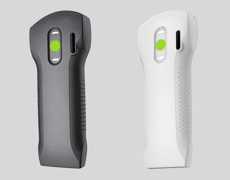
Quick reference: departments at a glance
| Department | Typical focused uses | Common probe array | Why portable fits |
|---|---|---|---|
| Emergency medicine | Focused trauma assessment for free fluid, focused cardiac views, lung assessment, vascular access | Convex + phased, or 3-in-1 | Minute-level decisions at the bedside |
| Critical care / ICU | Volume status trends, pleural and peritoneal assessment, guidance for lines and drainage | Convex + linear | Repeated imaging through a shift |
| Anesthesia & pain | Nerve block and regional anaesthesia localization | High-frequency linear | Needed at the moment of the procedure |
| Vascular access teams | Vein mapping, cannulation guidance | High-frequency linear | Goes to any ward on demand |
| Cardiology (clinic) | Focused cardiac windows, pericardial assessment | Phased array | Between consulting rooms |
| Obstetrics & gynecology clinic | Obstetric and pelvic clinic views | Convex (some use endocavity) | High patient throughput across rooms |
| Urology | Bladder volume, renal views, prostate assessment | Convex + biplane rectal | Bedside bladder scanning; steady clinic flow |
| MSK / sports / rehabilitation | Tendon, muscle and superficial structures | Linear or high-frequency linear | Sideline, clinic and bedside |
| Primary care | Triage and focused clinic assessment | Convex or 3-in-1 | Broad case mix with one device |
| Prehospital & EMS | Transport triage, focused assessment | Convex or 3-in-1 | Battery powered, zero setup |
| Veterinary | Companion animal and livestock reproduction, backfat measurement | Convex / linear / rectal by species | Farm, field and mobile work |
The departments that use it most

Emergency medicine
This is usually the first department to adopt. The questions are binary and urgent: is there free fluid after trauma, are there pericardial collections, how is global cardiac function, do we see lung sliding, where is the vein for this line. Because several body regions get covered in one shift, many ED teams start with a three-in-one probe (convex + linear + phased) rather than three separate probes. Our emergency POCUS guide walks through the workflow in detail.
Critical care and ICU
Here the value is repetition. Volume status, pleural collections and the position of lines or drains are checked repeatedly, so a device that lives in the unit removes a dependency on scheduling. Convex covers the abdominal and pleural questions; linear handles the superficial and vessel work. See the critical care application page for typical use patterns.
Anesthesia and pain medicine
The defining feature is that imaging happens during the procedure, not before it. Regional and nerve blocks rely on seeing target nerves and surrounding structures in real time, which makes a high-frequency linear probe the usual choice, with the device positioned next to the operator. Refer to the anesthesiology application page.
Vascular access teams
Vessel mapping and cannulation guidance sit close to the surface, so this work belongs to a high-frequency linear probe. Because these teams are called to many wards, portability is the point — the device travels rather than the patient. More on the vascular use case is on the vascular application page.
Cardiology clinics
Cardiac windows require a small footprint probe that can reach between the ribs, which is why a phased array probe is the standard here, often complemented by spectral Doppler for flow assessment. In outpatient practice the benefit is moving between consulting rooms quickly. See the cardiology application page.
Obstetrics and gynecology clinics
Clinic sessions are high-throughput and room-based, so a convex probe covering obstetric and pelvic views typically anchors this setting; some practices add endocavity capability. Portability here is about keeping pace across several rooms rather than about extreme ruggedness. Details on the OB/GYN application page.
Urology
Two different tasks live here. Bladder work benefits enormously from a handheld device — measuring bladder volume at the bedside without moving the patient is a classic use case, see our bladder scanner range. Prostate-related assessment uses a biplane rectal probe, which gives two perpendicular planes from one position. More context on the urology application page.
Musculoskeletal, sports medicine and rehabilitation
Tendons, muscle layers and superficial soft tissue sit within a centimetre or two of the skin, so linear — and for small joints, higher-frequency linear — is the working array. These settings often run clinics, sidelines and treatment rooms rather than a fixed imaging suite. See the MSK application page.
Also common: primary care, prehospital and veterinary
Primary care typically sees the widest case mix with one device, which is why a convex or three-in-one probe is often the pragmatic choice; see the primary care page. Prehospital and EMS need battery operation and the absence of any setup delay. Veterinary work spans companion animals to livestock reproduction and backfat measurement, with the array chosen by species — see the veterinary ultrasound range.
What multi-department buyers get wrong
Buying one "best" device for everybody. Probe-array requirements genuinely differ. A pure linear device is excellent for blocks and useless for abdominal free fluid.
Ignoring recurring software cost. A cheap device plus annual fees across many units can exceed the hardware budget over a few years.
Training last. Because image quality depends heavily on the operator, budget for training alongside hardware.
Forgetting about form factor. Some departments need true pocket carry; others are fine with a laptop-format unit. Our handheld vs laptop vs cart comparison covers that trade-off.
A sensible way to standardise
Map departments to probe arrays first, then decide how many devices each department needs — start from our types of ultrasound probes guide.
Pick one supplier across tiers so training, accessories and service are shared.
Deploy a mix: three-in-one probes for general and emergency use, dedicated linear or phased probes for high-volume specialist departments.
Verify quality-system credentials — see how to verify a handheld ultrasound manufacturer.
Confirm software licensing and total cost before scaling, and check the 2026 handheld ultrasound buyer's guide for spec comparison.
Planning units for several departments?
Browse SonoStar wireless handheld probes and the portable laptop range, or request a department-by-department configuration proposal.
This guide is educational content about equipment categories and departmental workflow. It is not medical advice, a clinical protocol, or a diagnostic recommendation. References to observing, assessing or assisting describe common focused-use patterns and do not imply diagnostic performance. Portable ultrasound does not replace comprehensive imaging or specialist interpretation. Image quality and safety depend on operator training and local clinical governance. Verify all credentials, specifications and current terms with the supplier before purchase.
- Pre:Handheld Ultrasound with No Subscription Fee: The TCO Buyer's Guide
- Next:No more




 Home
 Product
 News
 Contact