


Linear-Array 3D Volume Imaging Handheld Ultrasound: What the Mechanical Sweep Adds
- 2026-11-21
- 42
- Guangzhou Sonostar Technologies Co., Limited
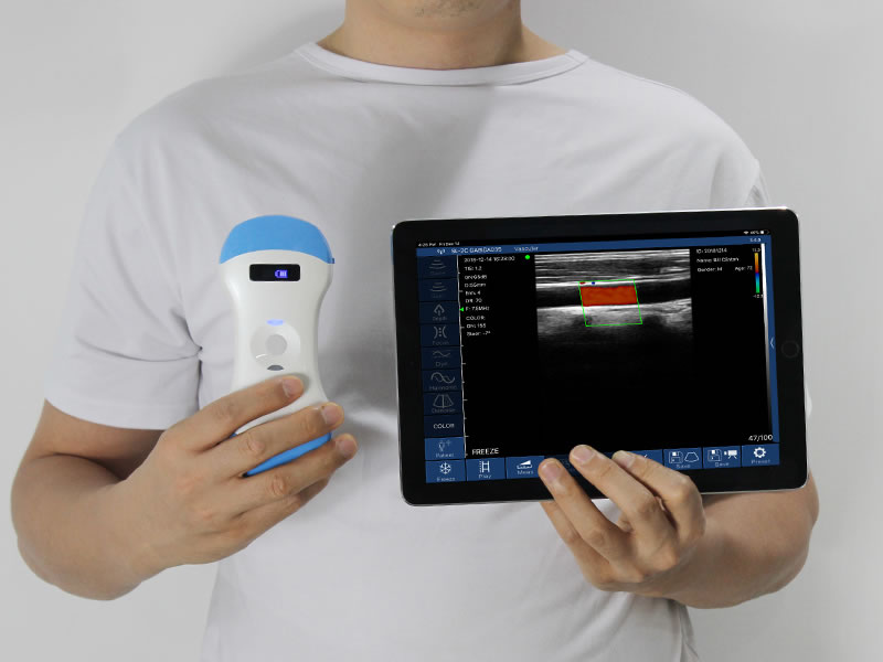
A linear-array 3D volume imaging handheld ultrasound builds a volume of shallow tissue rather than a slice: the array fires as a mechanism inside the head swings or rotates, and the resulting dataset renders on a phone or tablet. This is not a smaller version of the convex 3D head — the imaging geometry differs, and so does the job. It works at 2 to 20 MHz over a short depth range, which makes it a tool for thyroid, breast, musculoskeletal work and vascular access, and it changes what a superficial examination can document.
Why a linear array needs a sweep
A linear array images a plane. Elements fire in sequence along the aperture, and the beam stays perpendicular to the skin, which is why the image is rectangular and the resolution is even from top to bottom. That geometry is the reason linear probes are trusted for shallow structures.
A plane gives no volume. To build one, either the operator sweeps by hand or the head does the sweeping. Hand-sweeping is free and it is also unrepeatable: the geometry depends on wrist speed and angle, and two operators produce two different volumes. A mechanical sweep inside the head fixes the geometry, so a rendered volume is reproducible and a second reader can re-slice it without re-scanning the patient.
The trade is frame rate and stillness. A moving mechanism needs time to finish a sweep, so a volume takes longer than one frame; and both patient movement and hand tremor blur it. In a thin neck or a breast, that means a steady hold for a few seconds — a trainable skill, not a trivial one.

What the volume adds in superficial work
Thyroid. Three orthogonal planes from one dataset show a nodule's margin against the capsule, its relationship to the trachea and the carotid, and its craniocaudal extent without three separate hand positions. For documentation and for a second opinion, that is the practical gain.
Breast. Margin, retraction and the distance to skin and pectoral fascia read differently in a rendered volume than in a single frame, which helps a clinician describe a lesion to a surgeon or a colleague.
Musculoskeletal. A tendon or nerve is a structure with length, so tracking it through a volume answers questions a single slice cannot: where the thickening begins, whether the course deviates, how a needle sits relative to the sheath along its path.
Vascular access and blocks. A stored volume can be cut in-plane and out-of-plane after the puncture, which is useful for teaching and for reviewing a difficult block. The needle is still guided in real time on the 2D view.
Where it fits clinically
The applications follow the frequency range, not the marketing. Thyroid and breast clinics; pain and anaesthesia for superficial blocks; rehabilitation and orthopaedics for tendon, nerve and joint work; vascular access teams for PICC and dialysis fistula assessment; superficial lymph node and skin lesion assessment; small-parts and paediatric superficial work. Veterinary practice uses the same shallow range.
The department that gains least is the one that needs depth. A linear-array 3D volume imaging handheld ultrasound images the top few centimetres well and says nothing useful about a deep abdominal organ; for that, a convex probe or a cart remains the instrument.

FAQ
How does linear 3D differ from the convex 3D probe? Depth and resolution. The convex head reaches 90 to 305 mm at 3.2 and 4.0 MHz for abdominal and obstetric work; the linear head works at 2 to 20 MHz over a shallow field for thyroid, breast, musculoskeletal and vascular use.
Can it replace a cart system for thyroid or breast studies? No. It gives a clinician a bedside view, a reproducible volume and a shareable file. Formal studies, measurements and signed reports stay with the cart and the sonographer.
Does the sweep require the patient to hold still? Yes, for a few seconds. Patient movement and hand tremor both blur a volume, which is why the technique is taught before the device is deployed.
As a professional manufacturer, SonoStarMed provides high-quality mini color Doppler ultrasound with complete international certifications such as CE, FDA and ISO13485. Custom OEM and ODM services are offered for global distributors and medical institutions. Every device undergoes strict quality inspection before delivery to guarantee stable imaging quality and long service life.For any related needs or specific inquiries, please feel free to communicate: 18126862475.
- Pre:Handheld Ultrasound: Enabling Point-of-Care Imaging at the Bedside
- Next:No more



 Home
 Product
 News
 Contact